
硅材料透射电镜样品制备研究
硅材料透射电镜样品制备研究(含任务书,开题报告,外文翻译,毕业论文10900字,答辩PPT)
摘要
本实验通过对硅材料透射电镜样品的制备,全面了解透射电镜样品的制备过程,熟知硅材料透射电镜样品制备的各个环节,并且能熟练的操作该过程中所需要的设备和仪器。制样过程主要包括圆片切割、抛光、凹坑、离子减薄等步骤,各个步骤都会对所制造样品的质量好坏有重大的影响,掌握和控制好每一步的实验条件和具体操作至关重要。其次,全面的了解透射电镜的工作原理并且能够利用透射电镜对所制备的样品作一个全面的分析和研究,通过观察和分析研究透射电镜所拍摄到的样品图片可以了解到样品的薄区面积并且判断出所制透射电镜样品质量的好坏,最后,再对所制样品的差异加以分析和研究,进而改进和完善实验方法和实验步骤,提高制造硅材料的透射电镜样品的效率。
关键词:透射电镜;硅材料;圆片切割;离子减薄;样品质量
The preparation and investigation of TEM sample of silicon [资料来源:http://Doc163.com]
Abstract
We can have comprehensive understudied process and well-know all aspects of the TEM sample preparation. Besides, We can skilled operate the apparatus and equipment required for this process .because of preparation of the silicon material TEM sample in this experiment. This process main includes wafer cutting, polishing, pits, Ion milling and each steps will affect the quality of the sample. Therefore, It is very important of mastering and controlling every steps of the experimental conditions. Secondly, We should comprehensive understand the working principle of the transmission electron microscope and can use transmission electron microscopy to prepare a comprehensive analysis and research for samples. We can understand the area of the sample of thin area and the quality of the samples is good or bad according to observation and analysis pictures of TEM samples .At last, we can find the optimal experimental conditions and reagents by analysis of the differences of the samples, At the same time ,we can improve the experimental methods and steps to improve manufacturing efficiency of the TEM sample of silicon [版权所有:http://DOC163.com]
Keyword: TEM; Silicon material; Wafer cutting; Ion milling; The quality of the sample
[资料来源:http://Doc163.com]
目录
摘要 I
1 引言 1
1.1透射电镜的发展历程 1
1.2 制备透射电镜样品的意义 2
1.3透射电镜的使用简介 4
1.4透射电镜样品制备的大致流程和步骤 5
2实验方法 7
2.1实验设备及材料 7
2.2实验步骤 7
2.2.1切割 7
2.2.2抛光 9
2.2.3凹坑 10
2.2.4离子减薄 13
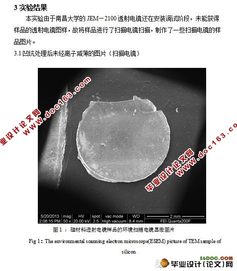
3实验结果 15
3.1凹坑处理后未经离子减薄的图片(扫描电镜) 15
3.2凹坑处理后又经离子减薄的图片(扫描电镜) 17
3.2.1 样品减薄时间长 17
3.2.2 样品减薄时间短 21
4结论 24
参考文献 26
致谢 28 [资料来源:http://doc163.com]